 |
Vaisblat A.V., Vesnin S.G., Konkin M.A., Lashchenkov A.V., Tihomirova N.N.
This paper discusses the method and results of the clinical trials that have been conducted among over 1500 patients.
Introduction
Presently the earlier detection of breast cancer is one of the most important problems. In several countries this disease is the main course of women death. In the United States one in every 9 women will experience breast cancer during her lifetime.
Specialists say that early detection of breast cancer by the clinical method is later biologically. Screening once in 12-24 months is not enough to detect fast growing breast cancer. Note that patients with fast growing breast cancer is a quarter of all breast cancer patients. …So it is expedient to use screening in conjunction with other non-invasive investigative methods [1].
Microwave radiometry [2-5] is based on measuring the intensity of natural electromagnetic radiation from a patient’s tissue. This intensity is proportional to the temperature of tissue. The change in temperature (thermal abnormality), that is a basis of the earlier detection of breast cancer, may be caused by increased cancer cell metabolism.
It should be noted that thermal changes precede to the anatomical changes that can be detected by traditional methods such as ultrasonography, mammography and palpation. Thus microwave radiometry is a very promising method for the breast cancer detection at an earlier stage. Also it should be noted that as microwave radiometry measures natural electromagnetic radiation from the patient’s tissue, it is absolutely harmless and safe both to the patients and to the medical personnel. So the method can be used successfully for screening for the monitoring treatment.
The specific heat generation in the tumor is proportional to the grow rate of the tumor. So fast growing tumors are “hotter” and they are more contrast in thermograms [6]. Thus microwave radiometry is an unique method that allows to detect first of all fast growing tumors. Using microwave radiometry (RTM-Diagnosis) in conjunction with other tradition methods allows to select patients with fast growing tumors [9].
The important feature of microwave radiometry is that it can distinguish proliferative mastopathy and fibroadenoma from non-proliferative mastopathy and fibroadenoma. So the method can select patients who risk to have breast cancer.
The main distinguish of microwave radiometry from well known infrared thermography is that the second allows to read and display the skin temperature, when microwave radiometry informs about temperature at a depth of several centimeters.
The first work discussing the use of microwave radiometry for the breast cancer detection was published in 1977 [3]. This subject was further discussed in the literature [6, 8, 10, 11]. At the same time the method has not been used widely in medicine practice.
Diagnostic System
In 1997 RES, Ltd. under the All-Russian Research Institute of Radio Engineering developed the RTM-01-RES microwave computer-based radiometer. The system includes a microwave sensor to invasively measure the temperature of internal tissue and a non-contact infrared sensor to the measure skin temperature. Information about the skin temperature allows to obtain more reliable results.
An abnormality can be detected at a depth of 3-7 cm, the accuracy of measuring the internal temperature is ±0.2 °C.
RTM-01 is a module null-radiometer with a sleeping scheme to compensate reflection between the object and the antenna. A wavelength of the radiometer is 26 cm. The scheme of the system is protected by the patent of the Russian Federation [7] and the device is approved to be used in medicine practice in Russia.
Radiation is received by a direct contact antenna that is set perpendicularly to the surface at the projection of the investigated organ. The device is safe, simple in operation and it has no regulators.
The main device specifications are the following:
Table 1
|
Items |
Specifications |
|
Thermal abnormality (i.e. a lower or higher temperature) is detected at a depth of, cm |
3 ‑7 (depending on water content tissue type) |
|
Accuracy of measuring the averaged internal temperature, when a temperature is 32 ‑ 38 °С, °С |
± 0,2 |
|
Time required for measuring internal temperature at a point, seconds |
10 |
|
Antenna diameter, mm |
39 |
|
Accuracy of measuring the skin temperature, °С |
± 0,2 |
|
Time required for measuring skin temperature at a point, when the temperature is 32 ‑ 38 °С, seconds |
1 |
|
Device mass, kg |
4 |
|
Power supply |
220 ± 22 Volt, 1 phase, 50‑60 Hz |
|
Power consumption, Watt |
20 |
The RTM-01 microwave computer based radiometer is shown in Fig. 1. The system includes a personal computer and a printer. The device is connected to a PC through a serial port. Results of RTM-diagnosis are shown in the monitor of the computer or printed out as a thermogram and temperature field on the projection of a investigated organ.
The advantage of the method is an expert computer system for the breast cancer detection. The expert system analyzes several parameters, including thermal asymmetry, dispersion of the temperature within the breast, etc.
|
Materials and Methods of Studies
The studies were held at Moscow oncological centers from December 1997 to September 1999. In total 1599 patients were examined. Some patients were examined several times, so 2154 examinations were performed.
According to CBE, mammography and histology the patients were divided into several disease groups (Table 2).
RTM-diagnosis is based on measuring the internal temperature and the skin temperature of breast. The procedure is made within 6-10 days of the menstrual cycle or on any day, if menopause. The patients lay on the back with their hands behind their head, in order to normalize the arrangement of the measured points and open axillary regions. Ten points on two breasts, including areola, centers of the quadrants, borders between the quadrants and axillary regions were measured. The measurement scheme is shown in Fig. 2. The skin temperature is measured by the same way.
Table 2
|
№ |
Disease |
Number of patients |
|
1. |
Health |
21 |
|
2. |
Fibrocystitis mastopathy |
181 |
|
3. |
Diffuse fibrocystitis mastopathy |
352 |
|
4. |
Fibroadenoma |
102 |
|
5 |
Node fibrocystitis mastopathy |
165 |
|
6. |
Cyst |
61 |
|
7. |
Fibrous fat involution |
55 |
|
8. |
Papilloma |
32 |
|
9. |
Mastitis |
49 |
|
10. |
Breast cancer |
316 |
|
11. |
The diagnosis of diseases is being exacted |
77 |
|
12. |
Other diseases |
188 |
|
Total: |
1599 |
Fig. 2. M

Measurement scheme of the breast

In some works discussing microwave radiometry temperature data
are displayed as a diagram, when the names of the measured points go along the
horizontal axis and the internal temperature values are along the vertical axis
(Fig. 3).
This method allows to analyze temperature differentials between corresponding points on the left and right breasts. However it is difficult to analyze the temperature at various locations on one entire breast by this method.
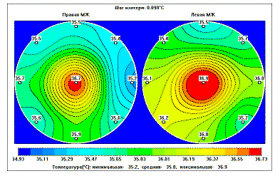
Therefore temperature data are also displayed as a temperature field, which is used in infrared thermography In the temperature field each temperature value is displayed by its own color on the monitor (Fig. 4).
In the temperature field cold areas of the breast are displayed by "cold" colours (i.e. blue) and hot ones are reflected by "warm" colours (red and pink).
Imaging thermal data as a internal temperature field show temperature abnormalities, that, in particularly, correspond to location of cancer.
 |
 |
Internal temperature depends on several factors. In particular, thin women with thyroid gland diseases have a higher internal temperature.
Fat women normally have a lower internal temperature. Increased dispersion is observed in women taking hormones, with fibrous-fat involution and during menstruation and ovulation.
Also lungs diseases may affect on the breast internal temperature. The measured internal temperature depends on the ambient temperature, so during examination an ambient temperature was 18-25 °C. Before the temperature reading procedure a patient should equilibrate to the ambient temperature. In order to exclude menstruation related changes in the internal temperature, the examinations were performed within 6 – 10 days of the menstrual cycle.
It is known that the temperature of the malignant tumor is 1-4 degree higher than the temperature of the surrounding tissue. So the temperature measured at the projection point of the tumor is higher than the temperature at the corresponding point on the health breast (point thermal asymmetry). In the temperature field this higher temperature is imaged as a red or yellow spot. Fast growing tumors have a higher temperature than slow growing ones, so they are more contrast in the thermograms.
Besides point thermal asymmetry normally breast cancer is accompanied by the increased nipple temperature of the diseased breast. 75% of breast cancer patients have a nipple temperature that is 0,5 degree higher than a normal temperature. For patients with the large breast, breast cancer are not always accompanied by the increased nipple temperature (especially, if a tumor locates on the periphery). Also breast cancer is accompanied by the increased differential between corresponding points on the right and left breasts and between points on one entire breast.
Different cancer types has different features that RTM-diagnosis detects. In particularly, ductal cancer is accompanied by the essentially increased nipple temperature (1,0 … 1,5°C) and a considerable thermal asymmetry (over 1°C) at one of points. Fig. 5 illustrates the internal temperature field of a patient who has ductal cancer of the left breast. In the temperature field a considerable thermal asymmetry and higher temperature of the entire left breast are reflected. This cancer type has exact RTM-features and there have not been mistakes in detecting it.

For comparison the temperature field of a health woman is shown
in Fig. 6.
Inflammatory cancer in contrast to ductal cancer is accompanied by the increase temperature at almost all points on the affected breast. Fig. 8 illustrates the temperature field of a patient with inflammatory cancer of the right breast.

RTM-features of inflammatory
cancer are similar to RTM-features of acute mastitis.
However the fact that the RTM‑diagnosis is harmless allows physicians
to order conservative treatment if there are suspicions of acute mastitis and
then repeat RTM‑diagnosis. This allows to compare results and analyse
dynamics.

Fig. 9a and 9b show a patient’s internal
temperature fields before and after conservative treatment. The second temperature
field confirms that there is no cancer.
 |
Fig. 9b demonstrates positive changes in internal temperature. (A fall in temperature is 20C). Thermal asymmetry has been also reduced.
 |
For women with higher breast temperature (group 3, table 3) breast cancer in 37% of patients is accompanied by inconsiderable thermal asymmetry (about 0,5°C). These patients are the most difficult cases for RTM-diagnosis.
Proliferative processes are accompanied by a local increase in the temperature, that is discussed below. So RTM-Diagnosis provides doctors with an unique ability to distinguish proliferative mastitis and fibroadenoma from non-proliferative mastitis and fibroadenoma. It is obvious that proliferative mastopathy is accompanied by less considerable thermal asymmetry than breast cancer, however exact parameters allowing to distinguish breast cancer from proliferative mastopathy and fibroadenoma have not been developed yet.
Computer Data Processing for Breast Cancer RTM-Diagnosis
Breast cancer is accompanied by different factors detected by RTM-Diagnosis. These factors depend on a patient’s age, breast structure, cancer type, ext. Besides, non-oncological diseases may be also accompanied by considerable thermal asymmetry. In particular, in 44% of patients with non-oncological breast diseases a thermal asymmetry exceeds 0.7°C. So it is important to distinguish breast cancer thermograms from non-oncological thermograms.
During investigations we analyzed a great number of thermograms and than selected six RTM-features of breast cancer. These parameters (RTM-features) were formalized.
1.
An increased maximal nipple temperature in comparison
with an average temperature of the breast.  , when
 =
=
 ,
ti – the temperature at eight points on the breast (1…8 in
Fig. 2).
,
ti – the temperature at eight points on the breast (1…8 in
Fig. 2).
2.
An increased nipple temperature differential
between right and left breasts  .
.
3.
An increased maximal temperature differential
between corresponding points on the left and right breasts  , the temperature
is compared at points 1-8, Fig 2.
, the temperature
is compared at points 1-8, Fig 2.
4.
An increased standard deviation of the temperature
at the corresponding points on the left and right breasts  , temperature
is compared by pair at 0…8 points shown in Fig.2.
, temperature
is compared by pair at 0…8 points shown in Fig.2.
5.
An increased standard deviation of the temperature
differentials at the corresponding points on the left and right breasts
 , when
 ,
,

6.
An increased standard deviation of the temperature dispersion in one entire
breast  , calculated
for a specific point 0…8 shown in Fig.2 for each breast and a larger value is
considered.
, calculated
for a specific point 0…8 shown in Fig.2 for each breast and a larger value is
considered.
Computer diagnosis is based on the following principals. All thermograms of verified breast cancer are stored in a computer. Then the software compares a current thermogram with these thermograms of verified breast cancer and analyzes whether the current thermogram (the thermogram of the diagnosed patient) is similar to these cancer thermograms basing on the RTM‑features described above. These RTM-features were represented as numerical values.
 |
The RTM‑features and their links are selected so as to points are compact in the diagram. This compact area is called a "risk area". changes in temperature, depending upon a patient age, was taken into consideration. If a patient’s parameters for all RTM‑features of breast cancer locate within the "risk area", this patient is similar to the cancer patients and the software, basing on processing results, gives a massage that the patient has breast cancer.
It should be noted that if a doctor diagnoses basing on only one parameter, a sensitivity for the method is 60-75%. If all RTM-features are considered the sensitivity is over 90% and the specificity is more than 75%.
The numerical data for each parameter are represented in Table 3.
1
group – women with lower temperature of the breast 
2
group – women who have temperature of  .
.
3
group – women with higher temperature of the breast  , where
 normal average
temperature.
normal average
temperature.
Table 3
|
Average temperature, degree |
|||||||
|
1 parameter |
2 parameter |
3 parameter |
4 parameter |
5 parameter |
6 parameter |
||
|
1 group |
cancer |
0.86 |
0.98 |
1.35 |
0.52 |
0.69 |
0.63 |
|
not cancer |
0.42 |
0.64 |
0.89 |
0.42 |
0.47 |
0.53 |
|
|
differential |
0.44 |
0.34 |
0.46 |
0.1 |
0.22 |
0.1 |
|
|
2 group |
cancer |
0.68 |
0.80 |
1.17 |
0.45 |
0.64 |
0.55 |
|
not cancer |
0.15 |
0.43 |
0.67 |
0.31 |
0.36 |
0.41 |
|
|
differential |
0.53 |
0.37 |
0.5 |
0.14 |
0.28 |
0.14 |
|
|
3 group |
cancer |
0.34 |
0.75 |
1.01 |
0.40 |
0.56 |
0.43 |
|
not cancer |
-0.10 |
0.36 |
0.53 |
0.25 |
0.28 |
0.31 |
|
|
differential |
0.44 |
0.39 |
0.48 |
0.15 |
0.28 |
0.07 |
|
The values of RTM-features lower as the average temperature increases. However a temperature differential between the cancer temperature and the normal temperature is almost constant.
The clinical trial at the Oncology Heath Center of the Health Committee has shown that RTM‑Diagnosis can distinguish proliferative mastopathy and fibroadenoma from non-proliferative mastopathy and fibroadenoma. Therefore it can select patients who may get cancer under unfavorable conditions. These patients should have an complex examination at specialised health centers. The results are represented in Table 4.
Table 4
|
Disease |
Number of examined patients |
RTM‑Diagnosis |
|
Mastopathy and fibroadenoma with proliferation |
11 |
9- Thermogram shows RTM-features of risk group 2- there are no RTM-features |
|
Mastopathy and fibroadenoma without proliferation |
18 |
3– Thermogram shows RTM-features of risk group 15– there are no RTM-features |
The table shows that RTM‑Diagnosis distinguishes proliferative mastopathy and fibroadenoma from non-proliferative mastopathy and fibroadenoma enough reliable. Thus one of the advantages of RTM‑Diagnosis is to select patients with fibroadenoma and mastopathy with proliferation. Other diagnostic techniques can not do this as they detect anatomical changes in the breast. RTM‑Diagnosis provides a doctor with information on active processes in the breast.
Clinical trials of RTM‑01‑RES
The clinical trials were held in 950 patients under the direction of leader Russian specialists at four Moscow medical centers. They are the following:
· The Branch #1 of the Mammology Health Center;
· The Municipal Hospital #40;
· The Russian Oncological Institute under the Science Center of the Russian Academy of Medicine Sciences;
· The Oncology Health Center of the Moscow Committee of Health.
The purpose of the clinical trials was to estimate the ability of the RTM‑01‑RES system to detect breast cancer and monitor the treatment of benign tumors. At the Oncology Health Center of the Moscow Committee of Health specialists estimated the ability of RTM‑01‑RES to select risk patients. The risk patients are patient that should be undergo complex diagnosis. The results of RTM‑diagnosis were compared with ultrasound and mammography.
RTM-diagnosis was made independently from clinical, x-ray and other examinations. The results of RTM-diagnosis were compared with results reported by histology. They were blind clinical trials (a doctor did not know results reported by other methods).
The results of the clinical trials are displayed in Fig. 11.

Fig. 11
Fig. 11 shows that all data are coordinated. The sensitivity for the method is 85-94%, the specificity is 75-80%, and the accuracy is 77-90%. These results are comparable with results of mammography.
Analyzing Cases of Earlier Breast Cancer Detection
In this section we discuss some cases of the earlier breast cancer detection. They includes cases when RTM-Diagnosis detects all RTM-features of breast cancer at the pre-clinical stage of tumor growth, when traditional methods (mammography, ultrasound, palpation and diagnostic puncture) can not detect breast cancer.
The following patients were diagnosed in the Brunch #1 of the Moscow Health Center.
|
1. Patient K., 58 year old, case history # 47204 |
|
|
02.03.98 – |
Mammography. Fibrocystitis mastopathy with fibrous character, scar changing. Directed to an oncologist. |
|
02.03.98 – |
CBE Complaint on hardening in the breast. Gynecology - health Objective.
Nipples and areolas without specific features. There is not nipple discharge.
The both breast with fibrocystitis mastopathy. The operation scar is hardened
at some parts (the patient said). Node mass is not detected. The region
lymph nodes are not enlarged. Directed to undergo RTM-diagnosis and Diagnosis: Diffuse fibrocystitis mastopathy |
|
04.03.98 - |
RTM-conclusion. The thermogram has features of left breast cancer locating in the upper inner quadrant. |
|
02.03.98 -10.03.98 |
Diagnostic puncture. Erythrocytes. Fat drops. |
|
08.07.98 - |
RTM-conclusion. The thermogram has features of left breast cancer locating in the upper inner quadrant. |
|
08.07.98 - |
CBE. The left breast has scar changing. Under the areola there is solid-elastic node neoplasm of 1 cm in diameter with an enough exact boundary. Region lymph nodes are not enlarged. Directed to undergo repeated mammography. Diagnosis: Node fibrocystitis mastopathy, Susp. Bl ? |
|
Diagnostic puncture. Unstructured masses, isolated leucocytes. |
|
|
16.07.98 - |
Repeated diagnostic puncture. Cancer cytogram. |
|
16.07.98 - |
Repeated mammography. Fibrocystitis mastopathy, at fibrous location there is node neoplasm of 1.5 cm in diameter with light boundary. Conclusion: Susp. Bl.. Final conclusion of the oncologist: Bl. Mam.sin. |
|
2. Patient M., 34 year old, case history # 43503 |
|
|
13.11.97 - |
CBE Complaint on pain in the left nipple for three months. Gynecology - health. Objective. The breast is developed proper. The left nipple is covered by a crust and hardened. There is serous nipple discharge. The breast with diffuse fibrocystitis mastopathy, the glandular tissue prevails. Node neoplasm is not detected. The region lymph nodes are not enlarged. Directed to undergo mammography and cytology. Diagnosis: Diffuse fibrocystitis mastopathy (adenose type) |
|
13.11.97 - |
Mammography. On the background of weak fibrocystitis mastopathy in the left breast under the areola there is a more solid mass without an exact boundary with many microcalcinators (probably sclerose type). In order to specify the nature of this solid mass, diagnostic puncture and short R – control (3 – 4 months) are required. |
 14.11.97
- 14.11.97
- |
RTM-conclusion. The thermogram has features of left breast cancer. |
|
13.11.97 – 19.11.97 |
Cytology. “Corneous” sand buckle, cells of plain epithelium with disparios of nucleuses, leucocytes, detritus. Inflammation cytogram. |
|
14.11.97 – 19.11.97 |
Diagnostic puncture. Objective. Inflammatory process in the left nipple at the regress stage. |
|
12.12.97 - |
RTM-conclusion. The thermogram has features of left breast cancer. The examination is performed on an incorrect day of the menstrual cycle, RTM-examination should be repeated. |
|
23.12.97 - |
Repeated CBE. Taken smear for cytology (discharged from the nipple). |
|
23.12.97 - |
Cytology “Corneous” sand buckle. |
|
06.01.98 - |
Repeated CBE. Scarification of the left nipple. |
|
06.01.98 - |
Cytology Erythrocytes, leucocyte, corneous” sand buckle, polymorphous cells of a malignant tumor. It is necessary to distinguish between cancer and black cancer. |
|
12.10.98 - |
RTM-conclusion. The thermogram has features of the left breast cancer. |
|
12.10.98 - |
Repeated CBE. Directed to an oncological center for operation. Diagnosis: Left nipple disease. |
|
Examinations in the Russian Oncological Science Center. Case history # 98/5198. The patient is in the hospital from 02.02.98 to 09.02.98 |
|
|
22.01.98 - |
RTM-conclusion. The thermogram has RTM-feature of left breast cancer. Cytology # 451 – cancer. Ductography – Padget cancer of the left beast. Histology # 1168. In the central part of the breast there is a node of 1.3 cm in diameter, on the projection of the nipple this node is 3 cm in diameter – Padget cancer spreading through the ducts to deep parts with infiltrating ductal cancer. In the axillary lymph nodes – hyperplasia. Final conclusion of the oncologist: Padget cancer of the left breast. IIа T2N0M0 |
|
3. Patient K., 41 year old, case history #45832 |
|
|
13.01.98 - |
Mammography. Complaint on pricking in the right nipple. Examination – diffuse lobulation, there is node movable elastic neoplasm on the border of outside quadrants of the right breast. Mammogram of 10.05.97 shows bilateral diffuse mastopathy. On the border of the outer quadrants of the right breast there is node neoplasm of 1.5 cm detected retromarnly. This mass has exact wavy boundary. The patient is directed to an oncologist. Conclusion: Cyst? (Fibroadenoma?) of the right breast. |
|
13.01.98 - |
CBE. Complain on pain and consolidation of the breast. Gynecology – cyst of right ovary. Objective. In the breast the inhomogeneity, lumpiness. In the right breast on the border of the outer quadrants (near them) there is a round movable mass of 1,5 cm in diameter with even boundary. Directed to undergo diagnostic puncture. Diagnosis: Fibroadenoma of the right breast. |
|
13.01.98 – 15.01.98 |
Diagnostic puncture. There is a few information – erythrocytes, isolated cells of cubic epithelium. Isolated cells of stroma, hemosidrofagy. |
|
15.01.98 - |
Repeated CBE. In the breast – st. Idem. The operation is postponed. Directed to 1. Ultrasound 2. RTM-Diagnosis on a correct cycle day. |
|
06.05.98 - |
RTM-conclusion. Risk group. The repeated examination is required. |
 17.11.98
- 17.11.98
- |
Directed to a hospital. Final conclusion of the oncologist: Fibroadenoma of the right breast. |
|
|
From the history case #35208. |
||
|
18.12.98 |
Performed sector resection of the right breast. |
|
|
Histology # 1021149 – Fibroadenoma of the breast. From the epicrisis of the history case #359. The patient K., 41 year old from 1.99 to 18.1.99 was treated in mammology department. She had right breast cancer T2N0M0 2a stage. The diagnosis was made by histology after sector resection of the right breast 18.12.98. Histology # 102150/153 – tumor of 2.8x2.0x1.5 cm. The mucous cancer. The mucous component prevails, with minimal number of parenchymatous elements. |
||
|
6.1.99 radical resection of the right breast is performed. Histology # 695-711 – nidal fibrosclerosis without defect of the tissue. In the lymph nodes of the axillary cellular tissue there is lymphoid reactive hyperplasia. After the operation the patient was consulted by a radiologist. Recommendations: distance gamma therapy for four weeks. 15.1.99 the radiation began. The post operation period is normal. The patient left hospital in satisfactory condition with stitches. |
||
Women included in the group of the earlier breast cancer detection (14 patients) have tumors of 0.5 to 3-4 cm in diameter. A time between RTM-diagnosis conclusion on breast cancer and disease verification is 1 – 7 months. For this period, if a tumor doubling time is 35 – 110 days [1], minimally volume of a tumor can increase 2-4 times, or its size increases by 30-60 percent.
Thus some of presently false-positive RTM-conclusions may with time become true-positive ones.
At present patients in the risk group have repeated RTM-examinations. Basing on obtained data we can believe that cancer verification continues.
Monitoring of Treatment
As RTM-Diagnosis is harmless, it can be used to monitor the treatment of various mastopathy types.

Dishormone hyperplasia is accompanied by changing in thermal
activity of breast tissue. So the temperature field of the patient is changed.
As a result of a specific treatment the temperature field may be normalized.
It confirms that the chosen treatment is proper. The literature [12] discusses
various treatments of dishormone breast diseases and shows that RTM-Diagnosis
is a sensible and effective method for monitoring the treatment of these diseases.
In contrast to breast cancer that is accompanied by higher temperature at cancer
location, benign tumors in some cases are accompanied by lower temperature at
some parts of the breast.
Basing on the temperature field a doctor can not verify a disease (e.g. distinguish fibrous from fibroadenoma or node mastopathy), as all these diseases are characterized by an uneven temperature field. As a result of the treatment the temperature field is normalized that confirms a proper treatment. Figures below illustrate two examples of using RTM-Diagnosis for the monitoring of the treatment.
Resume and Some Perspectives

The clinical trials and experience of using
RTM‑01‑RES at the leader oncological centers have shown that microwave
radiometry is an effective method for the earlier breast cancer detection. As
it is harmless, it allows to select patients with abnormal thermograms at an
earlier stage. These patients should undergo the complex breast examination.
The sensitivity for the method is comparable with the sensitivity for mammography
and ultrasound. The method is very promising to be used for diagnosing young
women, as mammography is not effective for this group. Also microwave radiometry
is useful for monitoring the treatment of benign diseases.
Mammology is not a single area where microwave radiometry can be used. The system can be used in urology, gynecology, for diagnosis of thyroid diseases, ext.
RTM-01-RES allows to read and display the internal and skin temperature. The next step of developing microwave radiometry is designing multi-frequencies devices allowing to read and display temperature distribution along depth and also creating multi-channel systems, that allows to minimize temperature reading time and labour-intensiveness of the examination.
Literature
1. В.М. Моисеенко, В.Ф. Семиглазов Кинетические особенности роста рака молочной железы и их значение для раннего выявления опухоли. Маммология, № 3, 1997 стр. 3-12.
2. В.М.Поляков, А.С.Шмаленюк СВЧ- термография и перспективы ее развития. Электроника СВЧ, вып.8(1640) Москва 1991г.
3. Barrett A., Myers P.C., Sadowsky N.L. Dedection of breast cancer by microwave radiometre. Radio Sci.-1977.-Vol 12, №68-P.167-171.
4. Троицкий В.С. К теории контактных радиотермометрических измерений внутренней температуры тел. // Изв.вузов. Сер. Радиофизика. - 1981.- т.24,. № 9- с.1054.
5. Сборник трудов всесоюзной конференции “Методические вопросы определения температуры биологических объектов радиофизическими методами” (Звенигород - 84) М.1985.
6. Carr K.L. Microwave Radiometry: its Importance to the Detection of Cancer. IEEE MTT, vol. 37 № 12 Dec. 1989.
7. Вайсблат А.В. Медицинский радиотермометр. Патент РФ № 2082118 с приоритетом от 11 июля 1994г.
8. Рахлин В.Л., Алова С.Е. Радиотермометрия в диагностике патологии молочных желез, гениталий, предстательной железы и позвоночника. Препринт № 253, Горький, НИРФИ, 1988.
9. Бурдина Л.М., Вайсблат А.В., Веснин С.Г., Конкин М.А., Лащенков А.В., Наумкина Н.Г., Тихомирова Н.Н. Применение радиотермометрии для диагностики рака молочной железы - Маммология 1998г. №2 стр. 3-12.
10. Бурдина Л.М., Вайсблат А.В., Веснин С.Г., Тихомирова Н.Н. О возможности диагностики рака молочной железы путем измерения собственного электромагнитного излучения тканей (радиотермометрии) - Маммология 1997г. №2 стр. 17-22.11. Малыгин А.А. Радиотермометрия в диагностике заболеваний молочной железы. Диссертация на соискание ученой степени кандидата медицинских наук. Н.Новгород, 1993г.
12. Наумкина Н.Г. Новые подходы к диагностике и лечению фиброзно-кистозной болезни молочной железы. Диссертация на соискание ученой степени кандидата медицинских наук. Москва, 1999г.
www.resltd.ru © 2001 RES, Ltd. Created and Designed by V.I.
