Health
Ministry of Russian Federation Russian Medicine Academy of Professional Development
USE
OF RTM-01-RES INTEGRAL INTERNAL TEMPERATURE DIAGNOSTIC COMPUTER BASED RADIOMETER
FOR BREAST CANCER DIAGNOSIS
(DOCTOR’S
MANUAL)
Moscow 1999
This Doctor’s Manual covers
abilities of the RTM-01-RES integral internal temperature diagnostic computer
based radiometer to detect breast cancer and other breast diseases at their
earlier stages. The method is based on measuring natural electromagnetic radiation
emitted from a patient’s tissues. It allows to detect changes in breast temperature.
It detects a temperature differential as well as an absolute temperature. Also
Doctor’s Manual covers basics of radiometry, a breast examination method, RTM-features
of breast cancer with drafts. Doctor’s Manual is intended for radiologists,
oncologists, gynecologists, and surgeons.
Doctor’s Manual is prepared
by specialists of the Mammology Branch of the Clinical Radiology Department
of the Russian Medicine Academy of Professional Development and the Moscow Mammology
Health Center, Dr. Burdina L.M., Dr. Haylenko V.A., Dr. Kizaev E.B., Ph.D. Haylenko
V.A., pH. Legkov A.A., Ph.D. Pinhosevich E.G., Ph.D. Mustafin Ch. K., Ph.D.
Vaysblat A.V., Ph.D. Vesnin S.G. Tihomirova N.N.
Introduction
The earlier detection of
breast cancer is a major problem for the medical community. In some countries
breast cancer is the leading cause of the death for women. In the USA one in
every 10 women will experience breast cancer during her lifetime.
Palpation, mammography,
or ultrasonography detect anatomical changes in the breast. RTM-Diagnosis detects
physiological changes in the examined organ (temperature changes in some areas).
RTM-Diagnosis detects internal temperature fields of organs. The temperature
changes that may be caused by inflammation or increased cell metabolism, accompanying
degeneration of tissues, precede to the anatomical changes that can be detected
by palpation, mammography, or ultrasonography. Therefore RTM-Diagnosis has a
potential ability to detect cancer at its earlier stage.
The breast cancer lifetime
consists of preclinical and clinical phases. At the preclinical stage cancer
can not be detected by traditional methods.
The growth rate is represented
by a tumor “doubling time” (a time required for doubling mass or number of cells).
Despite that the DT varies widely, the tumor growth is always represented by
an exponential curve, i.e. the doubling time is a constant for a specific patient.
The specific heat generation
is inversely proportional to a DT, therefore the most dangerous tumors with
a short DT are “hotter”, and so they are detected by RTM-Diagnosis first of
all. Thus RTM-Diagnosis has a unique ability to detect fast growing tumors.
Using RTM-Diagnosis in conjunction with other diagnostic methods allows to select
patients with fast growing tumors (short DT). According to current data these
patients are a quarter of all breast cancer patients.
Method Benefits and Side
Effects
Benefits
RTM-Diagnosis can be performed
for patients of all ages and with widespread diseases.
Therefore the doctor can
perform an examination repeatedly, monitor the disease dynamics as well as effects
of radiation and drug therapies.
Side Effects
The method detects natural
electromagnetic radiation from the tissues, so it is absolutely harmless for
the patients as well as for the medical personnel. Therefore there are no side
effects.
Technical Basis of Method
The RTM-01 radiometer
is a null-radiometer with the slipping circuit for compensating reflection
between the antenna and the object. The device scheme is protected by the
patent #2082118 of the Russian Federation.
The Commission on Devices
and Technical Support of Oncology and Medical Radiology of the Committee on
New Medical Techniques of the Ministry of Public Health and Medical Industry
of the Russian Federation recommends the serial production and the use of
the radiometers, developed by RES Ltd., in medical practice. The refined name
of the radiometer is “ the RTM-01-RES computer based radiometer for measuring
the integral internal temperature of soft and bone tissues.”
Д –В. Code- 94 4125 0003.
Code – ОКДП (ОК 004-93) 3311222.
КЧ – 04.
Name – RTM-01 Medical
Radiometer
ID Number - ТУ 9441-001-39549185-98
(ДКГП.942232.001 ТУ).
The device is manufactured
and sold by RES, Ltd. (22, Bolshaya Pochtovaya, Moscow, 107082, Russia)
For receiving natural
electromagnetic radiation the antenna contacts the skin at a point-projection
of the examined organ or its part.
The device specifications
are the following:
|
Items
|
Specifications
|
| Thermal
abnormality (i.e. lower or higher temperature) is detected at depth
of, cm |
3
-7 (depending on water content tissues)
|
| Accuracy
of measuring the averaged internal temperature, when the temperature
is 32 - 38 ° С, ° С |
±
0,2
|
| Time
required for measuring internal temperature at a point, seconds |
15
|
| Antenna
diameter, mm |
39
|
| Accuracy
of measuring the skin temperature, ° С |
±
0,2
|
| Time
required for measuring skin temperature at a point, when the temperature
is 32 - 38 ° С, seconds |
1
|
| Skin
temperature diameter, mm |
12
|
| Device
mass, kg |
4
|
| Power
supply |
220
± 22 Volt, 1 phase, 50-60 Hz |
| Power
consumption, Watt |
20 |
The radiometer can be
used with a PC (fig. 1). RTM-01 is interfaced to a computer by RS-232.

Fig.
1.
The device set includes
the following items:
Internal temperature
sensor with antenna (ITS);
Data processing unit
(DPU);
Skin temperature sensor
(STS);
Cables.
If a PC is used the device
set includes the following extra items:
PC (SIЕMENS NIXDORF
SCENIC PRO D5 (C5));
Color printer, A4.
The simplified scheme
of the complete device set is illustrated in Fig. 2.
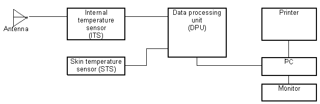
Fig. 2.
Device Operation.
The antenna contacts the
skin at a investigated point and receives natural electromagnetic radiation
as power that is transmitted to the ITS. In the ITS the signal is amplified
until it can be processed and converted to low frequencies.
After that the low frequency
signal representing information on the internal temperature is processed in
the DPU and the averaged temperature is displayed as a 3-digit number on the
temperature indicator located on the DPU.
The signal received by
the skin temperature sensor is transmitted to the DPU, where it is processed
in the same way.
The skin-internal temperature
switch is the sole controller on the device besides a power switch.
There are not external
regulators on the device in order for simplifying a doctor’s work.
The radiometer has some
hardware advantages. Due to its scheme RTM-01 measure temperature differentials
and absolute temperature with high accuracy. The device does not require calibration
testing.
The device scheme allows
to compensate reflection between the antenna and the bioobject.
The RTM-Diagnosis software
allows a doctor to:
Store and process patients
data, anamnesis and other additional information;
Automatically transfer
the internal and skin temperature data to the computer;
Display the measurement
results as a diagram;
Create internal temperature
fields;
Create and print out
an RTM-examination protocol including the patient’s data, anamnesis, a thermogram,
an internal temperature field, a conclusion on the possible disease, and
other data.
Also the RTM-Diagnosis
software includes a diagnostic expert system that compares the current patient’s
thermogram with the verified breast cancer thermograms and makes a conclusion.
The thermogram, where
the numbers of the points go along the horizontal axis and the internal temperature
values are along the vertical axis, represents actual data of the temperature
distribution. Imaging the data in such manner is not obvious enough, as the
measured points are displayed along a line, but in reality they may be located
along a circuit (breast) or other geometry, depending on the investigated
organ.
Obtained temperature data
may be displayed as an internal temperature field, on which measured points
are marked and isotherm lines are drawn through points with the same temperature.
In these fields temperature
abnormalities that, in particular, correspond to malignant tumors are displayed
well. (Fig. 5c).
For a gray scale image
sites with high temperature are displayed by light colors and sites with low
temperature are displayed by dark colors.
For a color scale image
sites with high and low temperature are displayed by warm (red) and cold (blue)
colors respectively.
The size of the temperature
is larger then the size of the malignant tumor reported by mammography, as
surrounding the tumor tissues are heated due to heat transferring and neuro-humoral
links. Therefore the resolution of the method does not contradict to sizes
of investigated thermal abnormalities.
Technical Requirements for
RTM-Examinations
RTM-Diagnosis must be conducted
in the diagnostic room complied with the following requirements:
The room area must be
10 – 18 m2. Lower floors are preferred, as the external noise level
is lower.
The room temperature must
be 20…24° C for reliable work of the device and patient convenience. When
the device is used in hot climate regions, air-conditioners must be set up
to provide this temperature.
Fluorescent lighting must
not be used in the room. Incandescent lamps must provide illumination of about
100 lux.
In the room there must
be four sockets grounded and complied with Class 1. The radiometer and the
PC must be connected to separated sockets. The distance between a PC and a
patient must be more than 1.5 m.
Method Description
The RTM-method is based
on measuring the internal tissue temperature of investigated organs, in particular,
the breast.
For this purpose natural
electromagnetic radiation from the internal tissues that is proportional to
the temperature of the tissues is measured. Infrared or liquid crystal thermographs
measure the skin temperature (epidermis). This temperature relates to the internal
temperature marginally.
Comments
A menstrual phase influences
on the temperature distribution in the breast. The examination must be performed
on the 6-9 day from the beginning of the menstrual period. Menopause women
can be examined at any time.
Palpation can influence
on the temperature distribution in the breasts, so it must follow RTM-Diagnosis.
The temperature distribution
is effected by hormone therapy, drugs, and contraceptives.
The work antenna surface
must be wiped with an alcohol tampon before every examination.
The examination technique
must not be varied and must comply with the recommendations defined below.
The radiometer may be used
in the following modes:
computer based mode;
manual mode (in this case
a PC is not used, obtained data are marked in the thermogram manually and
if it is necessary they may be input into a computer manually for analysis);
The breast temperature is
measured at the centers of the quadrants, at the borders between the quadrants,
at nipple points and lymph node points – a total of 20 points. (Fig. 4)
Examination
Scheme of Breast
0 – nipple
1 – border between upper
quadrants
2 – upper inner quadrant
3 – border between inner
quadrants
4 – lower inner quadrant
5 – border between lower
quadrants
6 – lower outer quadrant
7 – border between outer
quadrants
8 – upper outer quadrant
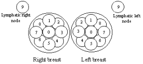
Fig. 4
To measure the temperature,
go through the following steps:
Turn on the radiometer and
the PC. Launch the RTM-Diagnosis software. The equipment must be heated for
15 minutes.
While typing the patient’s
general data (a surname, a history number, anamnesis), say some words about
the method to the patient, point out that it is absolutely harmless for her.
Especially it is necessary, if a patient is nervous and is afraid to be diagnosed
with cancer.
The patient must lay on
the back with her hands behind her head. The breasts are flattened and distribution
of the measured points is more convenient for performing the procedure.
For 20-30 seconds contact
the antenna work surface with the patient’ skin at the point of the solar plexus
projection in order to the antenna be heated to the patient skin temperature
and during further measurements the patient not feel cold that can affect a
measurement accuracy.
Contact the antenna work
surface with the right nipple. Do not press very much, but the entire antenna
surface must contact the skin (a visual control).
Substances for improving
antenna contact with the skin as they are used in ultrasonography are not required.
The internal temperature
sensor must be perpendicular to the surface, as obtained data depend on the
antenna slope angle.
Check to make sure that
a current measured point displayed on the monitor is the same that you are measuring
(right nipple).
In 10-15 minutes, once the
temperature gets a stable value, the sound signal and the scheme displayed on
the monitor will alert you.
Press the ЗАПИСЬ
(Record) button on the ITS or Enter on the keyboard.
Contact the antenna with
the left nipple and repeat the procedure.
The scheme displayed on
the monitor prompts a doctor a current measured point. Once you have measured
the first point on the right breast, measure the same point on the left breast,
and so on.
Once the measurement is
complete, review the thermogram. If it is necessary re-measured the temperature
at uncertain points.
To check the reliability
of the results measure the temperature at the right and left nipple repeatedly.
If the results differ from the first data to ± 0.5° C, repeat the whole procedure.
Obtained data can be displayed
on the monitor as
Temperature table (Fig.
5a)
Thermogram (Fig. 5b)
Internal temperature field
(Fig. 5c)
Risk group scheme (Fig.
5d) showing how much a current patient’s parameters are close to the risk areas
created basing on verified breast cancer thermograms.
Numeric estimate how much
a current patient is close to the risk group.
 |
| Fig.
5a |
|
|
| Fig.
5b |
|
|
|
Fig. 5c |
|
|
| Fig.
5d |
Fig 5
The data may be printed
out. The protocol of RTM-examination is represented in Attachment 1.
Breast cancer is accompanied
with the higher temperature due to increased metabolism of tumor cells.
There are the following
RTM-features of breast cancer:
Increased thermal differentials
between the corresponding sites on the left and right breasts.
Increased differential
between sites on the same breast.
Higher dispersion of the
temperature differential between the left and right breasts.
Higher standard deviation
of differentials between corresponding sites on the left and right breasts.
Differential between the
nipple sites on the left and right breast.
Higher nipple temperature
in the damaged breast in comparison with the average breast temperature. The
age-related changes must be considered.
All verified breast cancer
thermograms are stored in a computer. The RTM-features of breast cancer defined
above are represented by numeric values. Parameters of all patients with breast
cancer are displayed in six 2-D diagrams. Each patient is represented by a point.
The mathematical representation of RTM-features is selected so as to points
are compact in the diagram. This compact area is called a "risk area".
The average breast temperature goes along horizontal axis. Age-related changes
in temperature are considered. (Fig. 5d).
If a patient’s thermogram
for all RTM-features of breast cancer locates within the "risk area",
this patient is similar to the cancer patients and the software, basing on processing
results, gives a massage that the patient has breast cancer.
The reliability of these
results increases as data are being collected. The results processing that is
based on the six criteria makes the diagnostics more reliable.
Consider RTM-features of
some cancer types. Ductal cancer is accompanied with a considerable increase
in nipple temperature (1-1.5° C) as well as a significant thermal asymmetry
at one point. (Fig 6.).

Fig. 6
Inflammatory
cancer is accompanied with a 1-1.50 С increase in the temperature
of the most part of the damaged breast. (Fig. 7)

Fig. 7
The same conditions accompany
acute mastitis. However the fact that the method is harmless, if there is any
suspicion of this disease, the patient may be treated with drugs and then the
RTM-results obtained before and after treatment may be compared. (Fig. 8)

Before treatment

b) After
treatment
Fig. 8
RTM-features are more obvious,
when fibrous fat involution exists, as transmission loss is less and so a thermal
differential increases.
Thin patients have a higher
average temperature, so a thermal asymmetry (temperature differential) is less.
In these cases the RTM-Diagnostic software may help.
Making conclusion a doctor
should consider
Thermogram;
Internal temperature field
Diagnostic expert system
conclusion.
Method Benefits
Using new diagnostic methods,
in particular, radiometric methods in medicine in conjunction with the traditional
methods (clinical breast examination, mammography, ultrasonography, needle biopsy)
is very useful.
Clinical trials of the device
has been conducted at three medical centers of Moscow: the Branch #1 of the
Moscow Mammology Health Center, the Municipal Clinical Hospital and the Blohin
Oncology Science Center of Russian Science Academy of Medicine. The clinical
trials carried out in 900 patients show that the method has a high sensitivity
for breast cancer diagnosis (~90%), the method allows to repeat the procedure,
monitor the disease dynamics and choose an appropriated treatment. These features
are main advantages compared with mammograph-PC. The device has worked at the
Mammology Health Center as well as at leading Moscow Oncological Centers. For
this period 2000 patients have been examined.
The expert system allows
to estimate the conformity of a current patient’s thermogram to various verified
pathologic thermogram. The method sensitivity for breast cancer diagnosis is
90%, an accuracy is 84.8%, and a specificity is 78.6%.
The RTM-01-RES internal
tissue temperature computer-based radiometer is recommended for using in medicine
practice for screening at consulting and oncology rooms and at specialized oncology
and mammology centers for detection of breast cancer and the monitoring of treatment.
Attachment
1
RTM-Diagnosis Room Equipment
and Suppliers
|
№
|
Item
|
Number
|
Purpose
|
| 1. |
RTM-01
medical radiometer |
1 |
Perform
RTM-Diagnosis |
| 2. |
PC
and printer |
1 |
Process
results |
| 3. |
Screen |
1 |
Enclose
a patient |
| 4. |
Bed
|
2 |
For
a patient |
| 5. |
Table |
2 |
For
equipment and a doctor |
| 6. |
Chairs |
2 |
For
a doctor and a patient |
| 7. |
Peg
|
2 |
For
a doctor's smocks and a patient’s clothes |
| 8. |
Filing
cabinet |
1 |
For
documents |
| 9. |
Washbowl
with hot and cold water |
1 |
For
a doctor |
| 10. |
Room
thermometer 0 - 50° С |
1 |
Monitor
room temperature |
| 11. |
Pillow |
1 |
|
| 12. |
Pillow
case |
2 |
|
| 13. |
Sheet
|
2 |
|
| 14. |
Oil-cloth
medical, 1 m |
2 |
|
| 15. |
Garbage
can |
1 |
For
rubbish |
| 16. |
Alcohol
|
|
Disinfect
an antenna |
| 17. |
Cotton
wool or gauze |
|
Wipe
an antenna |
www.resltd.ru
© 2001 RES, Ltd. Created and Designed by V.I.


