
Doctor’s Manual
This Manual may be used by doctors who use or intend to use the medical radiometer for diagnosing diseases. Manual covers what place radiometry takes among diagnostic procedures, physical radiometry bases, features of the PTM-01-RES radiometer, and its structure. The diagnostics of oncological diseases is considered specially.
Radiometry is a new, developing area of disease diagnostics, especially it is helpful for the detection of oncological diseases.
The method is based on measuring the temperature of internal tissues by valuing their thermal radiation at microwave frequencies.
The radiometric method (RTM) has some advantages in comparison with traditional diagnostic methods. They are the following:
The main diagnostic means used for oncological disease diagnosis may be divided into three groups that are shown in fig.1.
Fig.1

Consider some features of these diagnostics. The first group includes physical examination methods. These methods are effective when the tumor is formed, and when it has a well delineated boundary. Unfortunately on the phase, when tumors have average size and so can be detected by physical methods (e.g. in average the tumor only of 1-2 cm can be detected by mammography) metastasis may occur.
Inevitable radiation load does not allow to conduct screening more than once every six months.
Physical methods have other features that restrict their abilities. For instance, in the mammology area the X-ray examination is not effective enough for young women as breast tissues are solid and so X-ray images are not contrast enough.
In the second group histology is the most reliable method. However it is applied during or after the operation.
Applying cancer markers is developing, however they are designed for diagnosing oncological diseases of limited organs group, and they are not always effective.
The third group includes thermal diagnostic methods. For the first time the temperature of the human body (oral temperature) was measured with the help of a mercurial thermometer (it had been just developed) in Germany in 1851. Since then temperature and its dynamics analysis has become one of the traditional diagnostic method. Noninvasive measuring internal organ temperatures began hundred years later due to developing night vision equipment during the Second World War.
During that procedure skin temperature that partially reflects measured internal organ temperatures. 20 years later (1972) the fist studies of internal tissue temperature measurement (tumors of mammary gland) were carried out. The measurement was based on receiving natural electromagnetic radiation at microwave frequencies. These studies were successful, as tissues are transparent enough for waves of this frequency range.
The distinctive feature of the thermal methods is that the methods are absolutely harmless and provide doctors with wide information.
Some special features of the methods are considered below. Note that temperature abnormalities caused by inflammation, increased cell proliferation at precancer stage or anaerobic glucose decomposition in the malignant tumor often precede anatomical changes that can be detected by physical diagnostic methods.
Detective ability of the thermal diagnostic methods is shown in fig. 2. The figure illustrates the results of the breast cancer detection that were obtained at the leading Moscow oncological center
Fig.2

Consider some well-known information on developing tumor growth and relationship between these data and the thermal diagnostic methods.
The tumor growth dynamics is characterized by the doubling time of a tumor (mass or the number of cells). The doubling time (DT) is a constant for a specific patient in spite of the fact that it varies widely (from 3 days to hundreds of days for different patients). Biological history of tumor growth may be divided into preclinical and clinical phases. The border between these phases is relative as it is determined by diagnostic equipment abilities. Therefore it is natural to apply the tumor growth behaviour studied for the clinical phase to the preclinical phase (doubling time is constant for a specific patient). The tumor growth is shown in fig. 3.
Fig. 3

When the doubling time is constant the tumor growth is represented by the exponential curve. It should be noted when the tumor mass doubles the tumor diameter increases times. Also tumors with short doubling time have a high specific heat generation (Watt/cm3). As when the tumor grows rapidly, energy consumption increases and so heat generation rises. This relationship is shown in fig. 4.
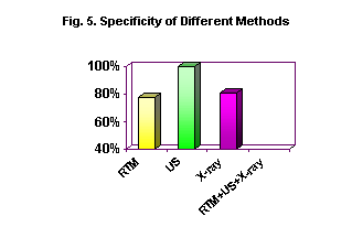

Fig. 4
Therefore most of dangerous tumors (tumors with short DT) can be detected by thermal methods first of all. It means that the thermal methods can select patients with rapidly growing tumors. According to current data these patients are a quarter of all breast cancer patients.
Consider transferring heat in a bioobject for better understanding the physic base of thermal diagnosis. As in any physical object, in the bioobject heat transfers from hotter areas to colder areas. There are three ways of heat distribution. They are heat transfer, convection and radiation. For the bioobject convection is represented by the circulation of the blood. They play their own role in thermal diagnostic methods. Fig. 5 illustrates heat transfer from internal organs to skin and then to ambient space, when there are no internal temperature abnormalities.
Fig. 5

In some parts of a human body the temperature is constant due to homeostasis. This temperature is approximately equal to the temperature measured in axial, oral and rectal areas (36.5° N – 37.0° N). The central nervous system, thorax organs, the abdominal area, and some parts of shoulder hips have a constant temperature.
When the room temperature is 20-25° C the skin temperature lowers to 32-33° C so there is a temperature gradient between the skin temperature and the internal temperature.
This temperature gradient and its changes, when a room temperature changes are shown in fig. 6.
Fig. 6

Radiation at infrared and microwave frequency is described by Planck’s radiation law. In accordance with it the level of emission is a function of both absolute temperature and frequency.
Fig. 7 shows emission intensity with respect to frequency, when a temperature is 310 K (33° N).
Fig. 7
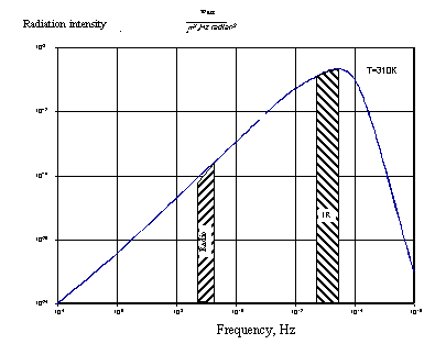
When temperatures are equal ones, which usually a bioobject has, the maximal radiation is at 1013 Hz frequency that corresponds to a wave of 10 micron, i.e. infrared radiation. The microwave frequencies used in radiometry (108 - 109 Hz) lay on the slope of Plank’s distribution and their radiation intensity is about 10 order less, that it is for infrared thermography. That determines device features for respective range.
Note that biotissues are not transparent for infrared radiation and it attenuates at a depth of several microns.
For microwave frequencies a transparency (attenuation) depends on water content of tissues. The tissues may be divided into two groups. The first group includes low water content tissues, which are represented by fat and bone. The attenuation of these tissues is low. It is 20-30% per depth centimeter.
Attenuation of skin and muscle (high water content tissues) is greater. It is about 50% per depth centimeter.
Consider some other features of thermal radiation. It is generated by chaotic movement of many molecules, so it is disordered (noise).
On the bioobject tissue – environment some radiation is reflected. It is governed by bioobject’s emittance according to Kirhgof’s law:
e = 1 - r, where
e - body emittance;
r - reflection coefficient.
Note that at infrared frequencies r is little (0.03 – 0.05), whereas at microwave frequencies r is 0.4 – 0.5. These features determine diagnostic device structure.
4. RTM - 01 – RES Diagnostic Set
Consider the RTM-01-RES diagnostic set as a sample of a medical radiometer. The RTM-01-RES is used for measuring the internal and skin temperatures. The measurement is based on receiving natural electromagnetic radiation at microwave frequencies and infrared frequencies respectively.
The simplified scheme of the device is shown in fig. 8. The diagnostic set consists of an antenna, an internal temperature sensor, a skin temperature sensor, a processing unit, a PC, and a printer.
Fig. 8
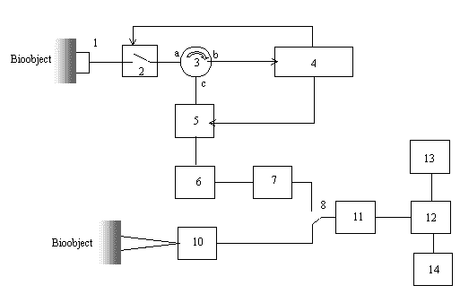
The internal temperature is measured by contacting the antenna (1) with a patient’s skin at the point of the investigated organ or its part projection.
Noise signal power at microwave frequencies received by the antenna is ? = ? K T B, where
K- the Boltzmann constant;
T – averaged internal tissue temperature (Kelvins);
B – microwave frequency band of the receiver (Hz).
? – emittance
.
When frequency band B is 100 MHz (108Hz) and the tissue temperature is 310K, this power is 4 o 10-13 watt.
The power is proportionate to a body temperature, so it can be measured in degrees, if other conditions are constant.
There is a switch (2) behind the antenna. It is switched from close to open position 1000 times per second.
When the switch is close the signal transfers through circulator legs a - c to the device radiometry region (4). When the switch (2) is open heated resistor (5) noise transmit to circular leg (c). This noise is reflected from the switch (2) and transmitted from circulator (3) legs a – c to the radiometer input (4). Signals are amplified and their power (temperature) values corresponding to close and open switch (2) positions are compared. Voltage that is proportional to the temperature differential between tissue and heated resistor (5) heats the resistor until these temperature are equal. Instead measuring the internal temperature we measure the temperature of the heated resistor, so that the device is simplified. There is a temperature-voltage converter. Voltage from the converter output achieves the mode shift (8) and then it achieves the analog-digital converter (11) that connects the device with the PC. The described scheme compensates reflection form skin surface. Fig. 9 illustrates this process.
Fig. 9
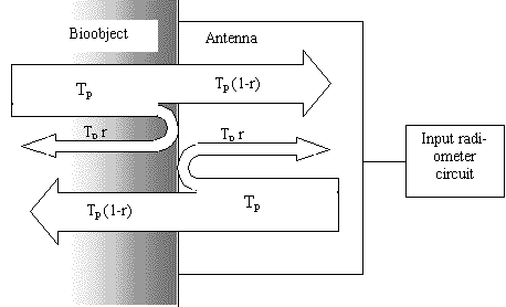
Noise power of a bioobject with temperature Op achieves the border between the bioobject surface and the antenna, then the antenna receives it reduced by 1-r according to Kirhgof’s low. Some noise Op x r is reflected and absorbed in the bioobject.
Noise with the temperature Op as mentioned above achieves the border from the receiver input. Some noise with temperature Op (1 - r) is absorbed in bioobject, and the rest of noise is reflected with the temperature Op x r. The sum of signal temperatures in the antenna input is Op.
So the reflection at the border is compensated even if bioobject are different.
The skin temperature is measured by a noncontact method. Intensity of radiation from a surface heated to absolute temperature T is
I = ? s (O 4 - O1 4)
where
? - heat-transfer coefficient;
s - Stephan – Boltzmann’s constant (5,67 o 10-8 watt / (m2 K4);
T1 – room temperature
The skin temperature sensor (10) consists of an optical system that forms a surveyed surface field, a mechanism that breaks ray flow from the compared body heated to the temperature O1 and a radiometric part.
A spot diameter on a patient’s skin depends on the distance between the antenna and skin.
When the distance is ten centimeters the diameter is 1.5 cm. When the distance is one centimeter the spot diameter is about 0.5 cm. These distances are set with help of special facilities.
The skin temperature and compared body temperature are compared with help of the mechanical breaker that operates 24 times in a second. The results of measurement are transmitted as direct voltage to the switch (8) and then to analog-digital converter (ADC) (11).
Besides the device the diagnostic set includes a PC processor (12), monitor (13) and a printer (14) that serve for
Temperature data are processed and can be displayed or printed as
For breast cancer diagnosis the special software is design. It allows to determine RTM-features of breast cancer, by analyzing a current patient’s data and the data of verified breast cancer patients.
5. Radiometer operation features
Consider some measurement features of the radiometer both at microwave and infrared frequencies, i.e. features of measuring skin and internal temperature.
The temperature that measured by internal temperature sensor consists of the following items:
Consider the relationship between temperatures measured by microwave and infrared sensors and true (thermodynamic) temperature distribution within internal tissues. Microwave antenna measures a brightness temperature. It reflects the internal temperature partially due to the following cause. If you image that investigated tissue is divided into thin layers that are parallel to skin, the emittance at microwave frequencies of each layer depends on physical temperature of the layer and radiation losses in it. Contribution of every layer to the brightness temperature depends on losses in the tissues placed from this layer to the skin.
Therefore the brightness temperature measured by the sensor that contacts the skin surface depends on thermal profile (T (z)) and electrophysic tissue properties, i.e. losses, as mentioned earlier. Fig. 10 illustrates the thermal profile when the thermal abnormality exists at the depth Z 0.
Fig. 10
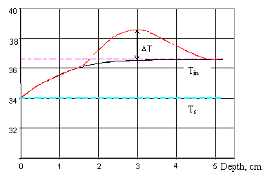
As the figure illustrates when there is no thermal abnormality the brightness temperature is a sum of skin temperature Ts (skin temperature) and the contribution of the temperature gradient. For typical differential Ti – Ts = 2,5 ° N (Ts is an internal temperature of the area, where it is considered constant) the contribution of the temperature gradient is 1 - 1,5° C and it is constant, when the skin temperature is constant. So internal temperature is always more than skin temperature. When the thermal abnormality occurs the picture is different.
Fig. 11 show computed brightness temperatures and skin temperatures for thermal abnormalities to be at deferent depths Zi. The following parameters are constant:
Fig. 11

Note that when thermal abnormalities are at little depth, the skin temperature may exceed the brightness temperature measured by the internal temperature sensor. This fact is helpful for diagnosis.
6. Measurement technique
Consider the technique of measuring the internal and skin temperatures. Reliable results can be obtained if the technique of measurement is not varied.
There are not any regulatory bodies on the RTM-01-RES except the power switch, ?AAEIAAO?EE (RADIOSENSOR), and AAO?EE OAIIA?AOO?U EI?E (SKIN TEMPERATURE SENSOR) buttons that switch operating modes. They are on the front panel of the processing unit. The mode indicator is on the temperature indicator panel.
When the device is turned on for the first time the ?AAEIAAO?EE (RADIOSENSOR) mode is set automatically.
Therefore the doctor can pay the whole its attention to measuring the patient’s temperature.
As mentioned earlier the room temperature affected both the skin and brightness temperatures. So the room temperature should be 20-25° C, and the best temperature range is 21-23° C.
There should not be draught in the room.
Before examination a patient’s skin temperature should become constant, for that the patient should adapt to the room temperature (for about 10 minutes.).
For measuring internal temperature there are no need for using any matter to improve contact between the antenna and the surface as it is used for ultrasonography. It is enough when the whole antenna surface contacts skin. The press on the surface is achieved due to the antenna weight that usually is vertical to the surface.
When the PC is used the measured point is prompted by a scheme displayed on the monitor. The CAIENU (RECORD) button allows temperature data to be stored automatically.
For improving measurement accuracy the measurement results are accumulated. This process last about 10 seconds. The indicator on the processing unit shows when the results become stable. Also you can see, when the accumulation process finished, on the scheme displayed on the monitor.
After that your should push the CAIENU (RECORD) button to store data. The temperature is stored when you set the button free.
There are two skin temperature measurement modes that are determined by the spot diameter on the skin. The temperature within the spot is averaged. As described earlier the spot diameter depends on the distance between the infrared sensor and skin. The distance is set by special facilities. When the distance is short (a diameter is 5 mm), the temperature of skin neoplasm is measured. When the distance is longer (a diameter is 15 mm), you can obtain additional data on internal temperature.
The accumulation time is mach less due to high signal intensity, and it is not more than one second.
The function of the CAIENU (RECORD) button is similar to one on the internal temperature sensor.
7. Examination methods
As mentioned earlier the measured temperature reflects the internal thermodynamic temperature partially. So for diagnosing abnormalities caused by different factors different diagnostic methods are used.
When temperature fields are analyzed the following facts should be considered. The abnormality size is always more than tumor size reported by the X-ray examination, ultrasonography and palpation due to the fact that the tissues surrounding a tumor or other source of high temperature are heated as a result of heat transfer.
Fig.12
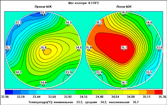
Fig. 12. Internal temperature field of a 57-year-old patient with left breast cancer (preclinical phase)
Fig. 13
Fig 14
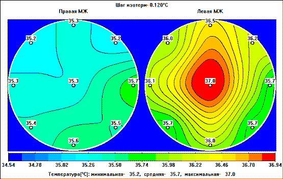
Fig.14. Internal temperature field of a 52-year-old patient with acute mastitis. Before treatment
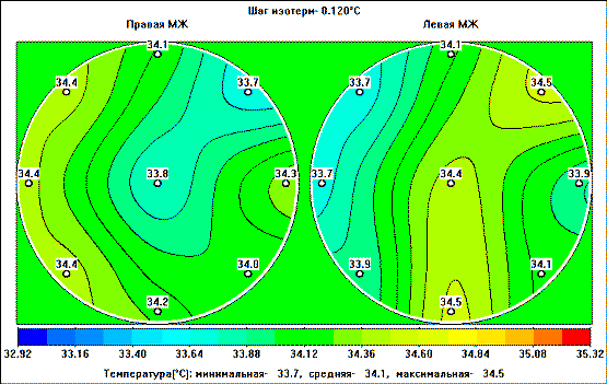
Fig.15
In conclusion note that the success of radiometric measurement mostly depends on desire of a doctor to obtain reliable data and it increases as the experience is gained.
Composed by
Alexander V. Vaisblat
Scietific Director & Head Researcher of RES, Ltd.
Manuscript
www.resltd.ru © 2001 RES, Ltd. Created and Designed by V.I.